At Khulna BNSB Eye Hospital, our mission is supported by a highly qualified and compassionate team of ophthalmologists and specialists, each with expertise in various subfields of eye care.
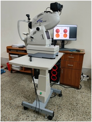
Fluorescein Angiography (FA) is a diagnostic test that helps ophthalmologists evaluate the blood circulation in the retina.
It also helps:
Rarely, an allergic reaction may occur, such as hives or difficulty breathing. In such cases, medical intervention is available.
Color Fundus Photography captures detailed color images of the retina using a specialized camera. This helps document and monitor retinal conditions over time
Before the procedure, pupils are dilated to improve visibility and allow a broader view of the retina.
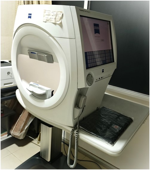
Visual Field Analysis measures your peripheral vision. It’s commonly used to assess conditions like glaucoma or detect visual impairment after a stroke.
You may blink normally and pause the test if needed.
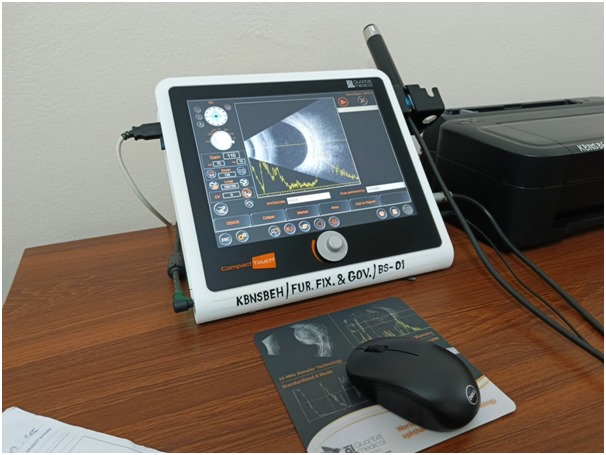
B-scan ultrasonography uses sound waves to produce an image of the eye’s internal structures. It is helpful in diagnosing:
It is also used before cataract surgery if the view of the eye’s interior is blocked.
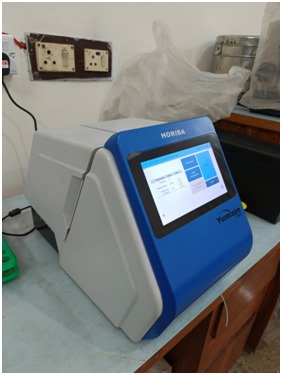
Pathology tests include analyses of blood, urine, stool, and tissue samples. These tests are often required before surgery or when diagnosing medical conditions. The results help guide clinical decisions and treatment plans.
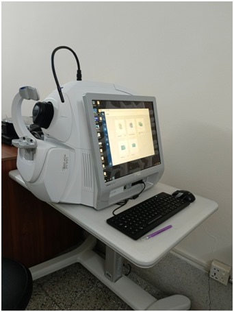
OCT is a non-invasive imaging technique that uses light waves to capture cross-sectional images of the retina.
This technology allows ophthalmologists to visualize and measure the thickness of each layer of the retina, aiding in the diagnosis and management of:
If dilated, your eyes may remain light-sensitive for a few hours afterward.
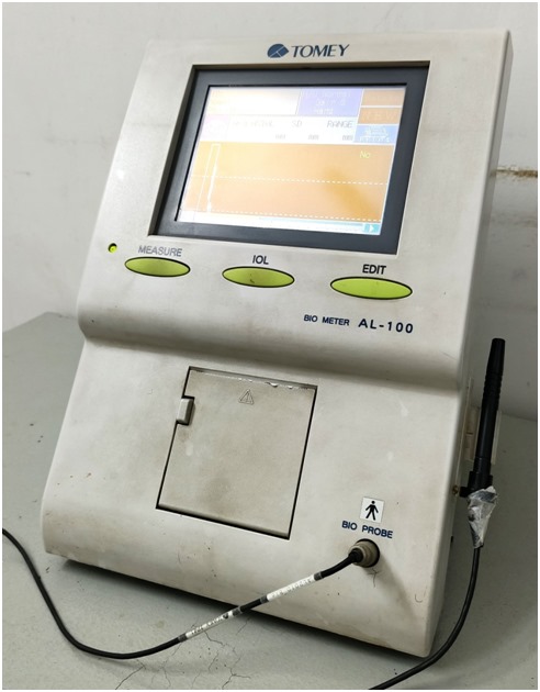
A-scan biometry is a diagnostic ultrasound technique used to measure the length of the eye, which is essential for calculating the power of the intraocular lens (IOL) to be implanted during cataract surgery.
These measurements are used to determine the ideal IOL power using specific formulas. Accurate biometry is crucial because even small measurement errors can significantly affect visual outcomes after surgery.
Even though modern optical biometry machines like the IOL Master are now common, A-scan is still widely used, especially when the optical pathway is blocked (e.g., dense cataracts). It remains a dependable tool in cataract surgery planning.
A retinal laser (also called laser photocoagulation) is a medical procedure used by eye doctors to treat diseases of the retina, which is the light-sensitive tissue at the back of your eye. The laser uses a focused beam of light to create small burns or controlled scars on the retina. These burns can seal leaking blood vessels, stop abnormal vessel growth, or secure the retina in place.
The YAG laser (short for Yttrium-Aluminum-Garnet laser) is a special type of laser commonly used in eye treatments, especially after cataract surgery. It delivers a quick, painless laser burst to clear cloudiness or to cut open membranes in the eye without any incision
Get to know our experienced medical professionals
Cardiologist
Neurologist
Pediatrician
Orthopedic Surgeon
Dermatologist
General Surgeon